The Life Cycle of Cryptosporidium parvum
 The
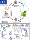
life cycle of C. parvum will be analyzed on this page. The
figure at the right helps to explain the different stages of
life for C. parvum and will be referred to as to help better
explain the life cycle of this organism. First, we can notice
that Cryptosporidium parvum has an zygotic life cycle. In a
zygotic life cycle, the organism has a unicellular diploid
stage. The unicellular, diploid stage is the oocysts, and is
looked for in determining if an environment has been effected
with or contains with C.parvum.
In stable conditions, such as in contaminated water or fecal
waste, oocysts of Cryptosporidium parvum are found
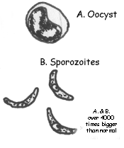
(Xiao et. al, 2004). An oocyst is a zygote of a parasitic organism
contained inside a fluid filled capsule or tough outer membrane.
The oocyst is the resting stage of an organism. An oocyst will
be what is referred to as stage 1 (as shown in figure 1.1).
Until an oocyst is ingested by a living organism, it will remain
in stage 1.
The second stage of C. parvum is to contaminate an
environment in which it might become in contact with its host
who will provide it nutrients to survive and and a protected
environment to reproduce. This may happen through poorly treated
pools, lakes, river, or public water. A case in Milwaukee, WI in
1993, 400,000 people became ill after they drank contaminated
public water (Xiao et. al, 2004). It is especially
threatening for people with
HIV
or AIDS. C. parvum also infects other animals, but most
commonly farm animals including
cows,
goats,
sheep,
and
pigs. Stage 3 of the C. parvum (as shown in figure 1.1)
is the stage at which an individual organism has ingested the
protozoan parasite. About 24
 hours after ingestion the oocysts
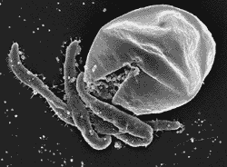
open to release sporophytes Stage 3-a. This process is known as
excystation or defined as an escape from a cyst (Borowski et
al., 2009). A sporophyte attacks a host cell (such as a human
cell) about five minutes* after excystation, and after 48 hours
all oocysts have opened. The sporophyte then develops into a
trophozoite; the stage (3-b) of life where a parasite acquires
nutrients from its host cell (Borowski et. al., 2009).
Asexual reproduction begins when the trophozoite stage
3-d,e,f divides to form two meront. Meronts are haploid cells of
C.parvum that reproduce asexually, and do so rapidly. This stage
has been observed as early as 24 hours after ingestion of the
oocyst . It has also been shown that meront I, which is the
first generation haploid stage of C. parvum, are smaller in size
and appear to have a thinner membrane then meront II, which is a
second generation haploid stage of C.parvum (Borowski
et al., 2009). When merozoites (unicellular haploid cells)
produced from meront II grow and develop, they form micro- or
macrogamonts, which are gametes preparing to undergo sexual
reproduction(stage 3-g,h). At this stage, the micro- or
macrogamonts are less connected to the host cell and more
extracellular (Borowski et. el., 2009.

Sexual reproduction occurs when a microgamont and macrogamont
undergo fertilization (stage 3-i). Together they form a zygote
and grow into an oocyst that can leave the host organism through
fetal waste or maintain the parasitic life cycle. This process
can take about 4 days (Borowski et. al., 2009). This means that
C. parvum can successfully live inside of a host, such as a
human for many days if they are not treated. Sexual reproduction
allows for C.parvum to exchange genes and evolve better to its
parasitic environment. These new oocysts (formed through sexual
reproduction) are believed to be largely responsible for the
life-threatening diseases of immunodeficient persons (Xiao et.
al, 2004).
For more information regarding the life cycle of C. parvum,
please check out this website below:
http://www.k-state.edu/parasitology/basicbio
*Time predictions are those provided by a study done on studied life cycles of C. parvum by H. Borowski, R.C.A. Thompson, T. Armstrong, and P. L. Clode. Look at references
Next is interactions, return to home, or to contacts.