Nutrition
Rhubarb is a plant, so therefore it is a primary producer, or an autotroph. This means that is produces its own food through a process called photosynthesis. The equation for photosynthesis is shown below.
+ 6H2O + 6CO2 --> C6H12O6 + 6O2
Photosynthesis starts with water (H2O), carbon dioxide (CO2), and energy from the sun that is taken in via the chloroplasts. Carbon dioxide and water come from the environment, but water can also come from the soil and be taken up through the roots. These starting materials react to produce sugar, specifically glucose (C6H12O6), and oxygen (O2), that came from the water. The glucose is used in cellular respiration to produce ATP, or energy. The oxygen is either used for respiration or released into the atmosphere.
Any excess food that rhubarb produces is stored in the roots as starch.
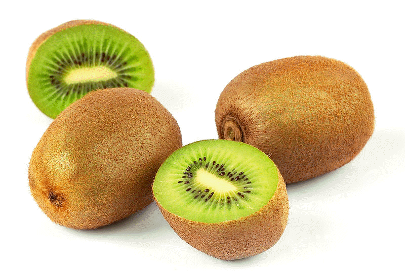
Other plants, such as the Kiwifruit, Actinidia deliciosa, and the Purple Passion Flower, Passiflora incarnata, get their food from this same process.
Rhubarb has a complex transport system made up of two vascular tissues, xylem and phloem, that distribute water and nutrients through the plant. Because rhubarb is a dicot, these transport tissues are arranged in a circular pattern around the stem, with xylem to the inside and phloem to the outside.
- The xylem is made up of dead cells, or vessel members, that form channels for the movement of water and minerals up the plant. Water is pulled upwards from the roots through the xylem.
- The phloem is made up of sieve-tube members and companion cells that work together to distribute nutrients through the plant. The solution that flows through the phloem is generally made up of sugars, amino acids, minerals, and hormones. This solution is pushed through the plant from source to sink. The source is usually the leaves, but it could also be storage roots. The sink is any tissue where the sugars are being used.
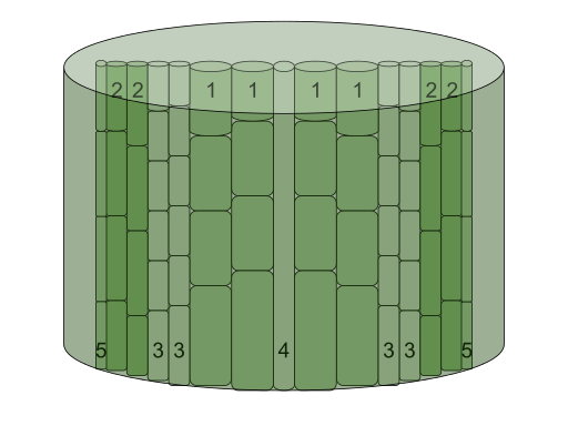
The image to the left shows a plant stem with xylem and phloem. Number 1 on the image shows the xylem, it is to the inside of the stem. Number 2 depicts the phloem, toward the outside of the stem. The companion cells of the phloem are depicted by number 5, and the remaining two areas are cambium (3) and pith (4).
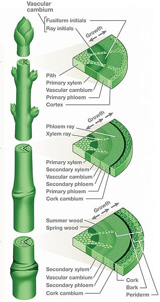
The image to the right shows a dicot plant stem with cross-sections showing the different tissues. Notice that the xylem and phloem are in a ring pattern all around the stem. Also, focus on where the xylem and phloem are located: xylem is to the inside and phloem is to the outside.
Continue on to Reproduction.
Go to Home. Go to Multiple Organisms. Go to UW-La Crosse.


